

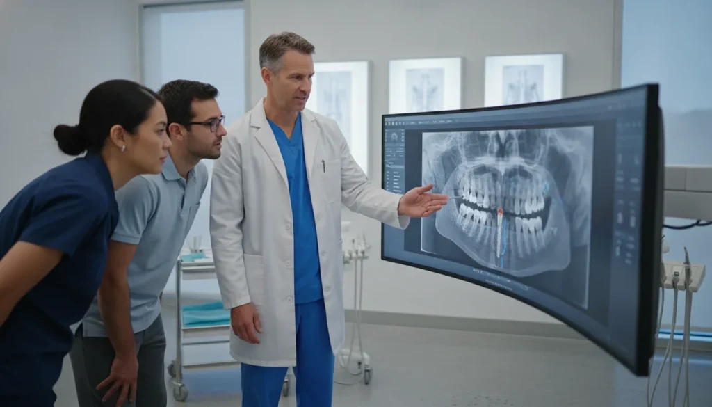
In an oral surgery practice, a radiograph is most useful when it is available at the moment the team needs it. If staff must search a separate imaging workstation, export files, or switch between applications during a patient visit, even a straightforward review can become slower and less consistent. The goal is not simply to capture high-quality panoramic, CBCT, or intraoral images. It is to connect those studies to the patient record and make them easy for authorized clinicians and staff to find.
Request a demonstration of MaxilloSoft’s oral surgery practice management software.
For practices evaluating carestream dental integration, the key question is whether the imaging workflow can move reliably into the practice management system or EMR without creating another disconnected archive. A well-designed connection can centralize radiographs, reduce repetitive file handling, and give the care team a clearer view of the patient’s history.
Carestream Dental integration can make radiographs easier to access in the EMR by connecting imaging studies with the patient’s centralized record. So staff spend less time toggling between separate systems during a patient visit.
That workflow starts with understanding the imaging technologies an oral surgery practice uses and how each fits into daily clinical and administrative work.
By Dr. Julius Hyatt, DDS, Founder and Oral Surgeon
How Carestream Dental Imaging Fits Into an Oral Surgery Practice
Carestream Dental can support an OMS imaging workflow by bringing panoramic views, CBCT studies, intraoral images, and imaging software into the broader practice management and EMR environment. So authorized clinicians can find the right study from the patient record instead of chasing files across separate applications.
Oral and maxillofacial surgery practices often need more than one type of image during a patient’s care journey. A panoramic study may support an initial overview, while a CBCT examination may provide a three-dimensional view for cases that require volumetric information. Intraoral sensors and cameras can add focused images to the patient’s clinical record. The practical question is not simply whether a device produces a useful image. It is whether the image can move reliably into the workflow where the OMS team documents, reviews, and communicates about care.
Carestream Dental’s portfolio includes extraoral imaging systems, such as panoramic and CBCT 3D platforms including the CS 8200 and CS 9600. It also includes intraoral digital sensors and cameras, such as the CS 6200 sensor and CS 1300 intraoral camera. The portfolio also includes imaging applications such as CS Imaging and CS 3D Imaging. These products are designed to serve different points in the imaging process, so practices can evaluate the equipment that matches their clinical and operational needs. Carestream Dental provides product and solution information for practices comparing these options.
Match the imaging source to the OMS workflow
A well-designed setup makes the source of an image less important than its accessibility in the patient’s record. When imaging data is centralized, the surgeon and referring provider can work from the same available study instead of relying on separate storage locations or manual handoffs. Carestream Dental’s imaging solutions can integrate with various oral surgery practice management systems, but the exact workflow depends on the equipment, software configuration, and receiving platform.
- Panoramic imaging: Supports broad extraoral views when the team needs an overview within the patient’s imaging history.
- CBCT imaging: Provides three-dimensional studies for OMS workflows that require volumetric information rather than a flat image.
- Digital sensors: Capture intraoral radiographs that can be associated with the appropriate patient record.
- Intraoral cameras: Add focused visual documentation that may complement radiographic studies and clinical notes.
Centralized radiographic data can help shorten the path from image capture to review and improve communication between surgeons and referring providers. It can also reduce the administrative effort involved in locating studies across disconnected applications. The goal is not to replace the imaging system. It is to make the resulting information easier to find and use within the OMS record.
For practices evaluating the connection between imaging equipment and their core system, the broader landscape of oral surgery software integrations is a useful place to start. Review which systems must exchange data, where staff need to view images, and how the workflow should behave from patient check-in through clinical documentation. Those requirements provide a more reliable basis for integration planning than choosing hardware in isolation.
Why View Radiographs Directly in Your EMR?
Viewing Carestream Dental radiographs directly inside the EMR gives an oral surgery team a single patient record and a single workflow. That means faster access to the imaging needed for clinical decisions, less time spent searching separate systems, and clearer communication across the care team during each patient visit.
When radiographs live in a separate imaging application, even a simple review can require staff to confirm the patient. Open another platform, search for the study, and then return to the practice-management system to document what happened. That repeated toggling may seem minor, but it adds friction throughout the day and makes it harder to keep the complete patient record in one place.
With the right integration, images associated with a patient can be available from the same EMR environment as demographics, surgical documentation, referrals, and scheduling information. This is especially useful for oral and maxillofacial surgery teams, where a patient may have multiple imaging studies across consultation, treatment planning, and follow-up.
Reduce administrative friction around every study
Centralization does more than make the interface feel cleaner. It reduces the number of steps required to locate a study and limits the need for staff to reconcile information across separate systems. Research on digital radiography in oral and maxillofacial imaging has linked implementation with improved workflow efficiency and broader access to radiographic data across dental systems (review the evidence on digital radiography).
- Centralized records: Keep patient information and related radiographs connected within the same clinical workflow.
- Less toggling: Reduce repeated movement between the imaging suite and the EMR or practice-management platform.
- Lower search burden: Give staff a clearer path to the study they need without hunting through disconnected applications.
- Consistent documentation: Review images while working in the patient record, then document the relevant workflow without changing context.
Teams evaluating this approach can also explore MaxilloSoft’s guide to viewing radiographs in your EMR for a broader look at imaging access and workflow design.
Support faster review and communication
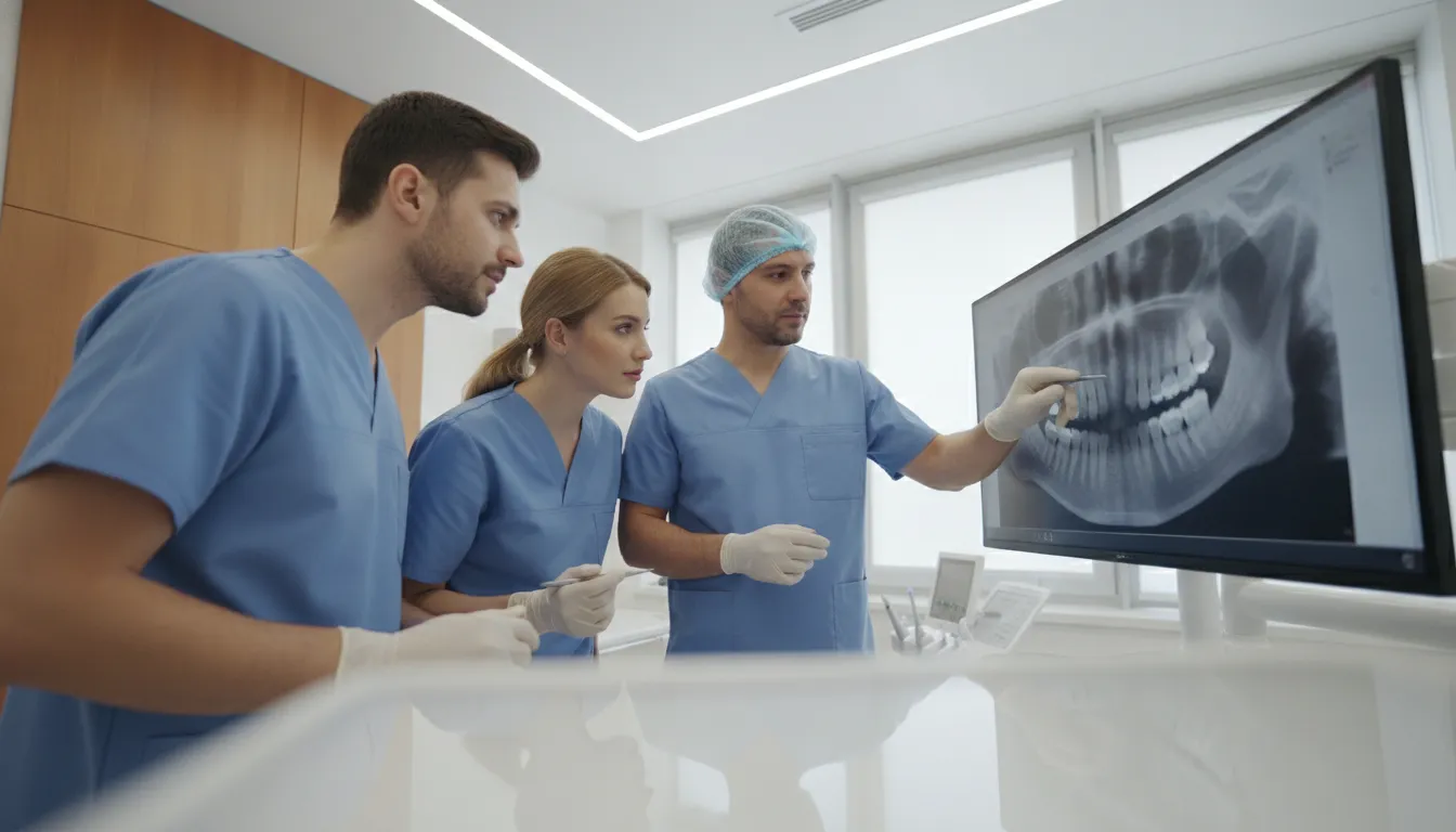
Centralized radiographic data can support faster diagnostic turnaround and more streamlined communication between dental surgeons and referring providers. The benefit is not a promise that every case will be resolved faster. Rather, the clinical team spends less time locating records and more time reviewing the information in context. When a referring provider asks about a study, staff can work from a more complete patient record instead of assembling details from multiple platforms.
For OMS practices, imaging access also needs to extend beyond routine two-dimensional radiographs. Consultations and treatment planning may involve volumetric CBCT studies, so the EMR workflow should accommodate the type of three-dimensional information the practice actually uses. The goal is a connected workflow that preserves access to the original imaging system’s capabilities while making relevant studies easier to reach from the patient record.
That is why integration planning should consider both the clinical workflow and the technical connection between systems. Standards such as DICOM are foundational to interoperability between imaging devices and electronic health record environments (see how DICOM supports imaging interoperability). Practices can then assess whether their EMR displays the right studies, maintains reliable patient matching, and gives authorized team members practical access without adding another disconnected destination.
For a broader framework, review MaxilloSoft’s guide to software integrations and evaluate how imaging access fits into the practice’s complete OMS workflow.
How Does Carestream Dental Integration Work in Practice?
A reliable imaging integration moves each study from the Carestream Dental environment into the correct patient record. Where authorized OMS team members can open and review it without sorting through disconnected files, repeating logins, or handling manual handoffs between separate imaging and EMR applications.
The practical goal is not simply to make two applications exchange data. It is to create a dependable path from image capture to patient documentation. That path should preserve the study’s patient association, retain the information needed to interpret it. And make the result available in the EMR or practice management workflow the team actually uses. Carestream Dental’s imaging stack can include specialized software such as CARESTREAM Image Suite, which the U.S. National Library of Medicine’s AccessGUDID identifies as a medical device used for diagnostic imaging support: see the device record.
- Establish the interchange: The imaging system and OMS software first need a supported method for exchanging studies. DICOM is foundational to interoperability between imaging devices and electronic health record systems, giving the applications a common structure for transmitting and interpreting medical images. In some environments, other healthcare data standards may support related exchange and reporting needs. The CDC describes standardized FHIR implementation as a bridge for data exchange between EHR systems and clinical registries, but it does not replace the imaging-specific role of DICOM: review the CDC guidance.
- Match the patient record: Before importing anything, confirm that the study belongs to the intended patient and that identifiers map correctly between systems. This checkpoint matters when a practice is bringing in existing panoramic radiographs, intraoral images, or CBCT volumes. A clear import policy should define which historical studies are included, how duplicates are handled, and who resolves exceptions.
- Import existing studies: Historical imaging can be migrated in a controlled batch or added as individual records, depending on the practice’s needs and the integration’s capabilities. CBCT volumes deserve particular attention because they contain more than a single flat image. The receiving system should preserve the study structure and make the appropriate viewer available, rather than reducing a volume to an isolated screenshot.
- Render within the EMR: Once the study is associated with the patient, the user should be able to open it from the patient record or an embedded imaging link. The experience should support the clinical workflow without forcing staff to search a separate application for every appointment. Centralized radiographic data has been associated with more efficient imaging workflows and broader access to dental radiographic information in oral and maxillofacial settings: read the peer-reviewed review.
- Apply role-based access: Configure permissions so surgeons, clinical staff, and administrative users receive the access appropriate to their responsibilities. Clinical users may need study viewing and documentation access, while front-office users may need only the patient and appointment context. Documenting these roles supports consistent onboarding and helps the practice review access when responsibilities change.
- Train, test, and support: Plan onboarding around real appointments, not just a technical demonstration. Test new captures, imported studies, CBCT viewing, patient matching, and common failure scenarios with representative users. Assign an internal owner for questions, record vendor support contacts, and agree on escalation steps before launch. A written workflow also makes it easier to compare oral surgery software options as the practice grows.
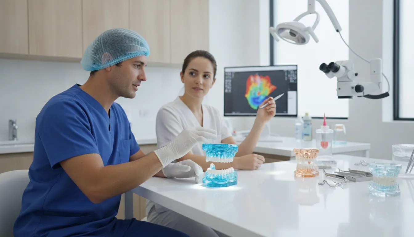
When these steps are treated as one operational workflow, integration becomes easier to validate and maintain. The practice can assess not only whether images technically transfer. But also whether the right people can find and use the right study at the right point in the patient journey.
What Should You Look For in OMS Software for Imaging?
The right OMS imaging workflow connects every study to the correct patient record, supports interoperable formats such as DICOM. And gives each team member access appropriate to their role, so clinicians can focus on care instead of managing multiple disconnected systems.
When evaluating practice management software, look beyond whether it can open a radiograph. The more important question is how imaging fits into the full surgical workflow. A practice may use Carestream Dental hardware and imaging applications. But the surrounding OMS software should make those studies easy to find, review, document, and share within established permissions.
Digital radiography has improved workflow efficiency and made dental radiographic data more accessible across an organization, according to a review of digital imaging in oral and maxillofacial practice. The underlying research supports evaluating imaging as part of a connected information system, rather than as an isolated workstation function.
| Capability | Native imaging portal in the EMR | Separate imaging viewer |
|---|---|---|
| Patient context | Images, notes, referrals, and surgical documentation stay connected to one patient record. | Staff may need to search for the patient again and reconcile information across systems. |
| DICOM import | Supports a defined path for receiving and rendering compatible imaging studies. | May depend on manual exports, local configuration, or a separate interface. |
| Team access | Role-based permissions can align access with clinical, administrative, and referral workflows. | Access rules may be managed separately from the practice record. |
| CBCT planning | Can provide a scalable foundation for volumetric studies as imaging needs grow. | May require additional software, training, and workflow coordination. |
Questions to ask during a software demonstration
Ask vendors to show the workflow using a realistic OMS scenario, not only a feature checklist. DICOM standards are foundational to interoperability between imaging devices and electronic health record systems, so confirm how studies are imported, matched, and rendered. This overview of DICOM interoperability illustrates why standards matter when systems need to exchange imaging data.
- Record continuity: Can staff open the image from the patient chart and document related notes without repeated searching?
- Permissions: Can administrators, surgeons, assistants, and referring partners receive the access appropriate to their role?
- Imaging range: Does the workflow support panoramic images, intraoral studies, and CBCT volumetric planning as the practice evolves?
- Onboarding: Who helps migrate existing studies, configure the interface, and train the team?
- Support: Is there a clear process for resolving import or display issues after implementation?
Centralized radiographic data can support faster diagnostic turnaround and clearer communication between dental surgeons and referring providers. Carestream Dental also offers imaging software such as Image Suite, which is identified as a diagnostic imaging support device in the AccessGUDID database. The best choice is therefore not simply the viewer with the most features. It is the OMS system that keeps imaging usable, secure, and connected to the work your team already performs. Review the broader integration approach before comparing individual products.
Frequently Asked Questions
How does Carestream Dental imaging integrate with practice management systems?
Integration connects the imaging workflow with the patient’s practice management or EMR record. Depending on the systems and configuration, staff can associate new studies with the correct patient, open imaging from the patient chart, and avoid switching between unrelated applications. Confirm supported interfaces, user permissions, and the implementation process with both vendors before deployment.
Can I view Carestream Dental radiographs directly in my EMR software?
Yes, when the EMR supports the required imaging connection and the integration is configured correctly. Staff can open radiographs from the patient record instead of searching for them in a separate imaging application. For oral surgery practices, confirm that the workflow accommodates both standard radiographs and larger 3D or CBCT studies when those are part of your clinical process.
What imaging systems does Carestream Dental offer?
Carestream Dental offers a range of extraoral and intraoral imaging products, including panoramic systems, 3D CBCT systems, digital intraoral sensors, and intraoral cameras. Its software tools support viewing and managing imaging studies. The right integration approach depends on the specific device, imaging software version, and interface supported by your EMR.
What should a practice plan before connecting imaging to its EMR?
Plan for interface configuration, patient matching, user access, existing-study migration, and staff training. Establish who will troubleshoot an unlinked or unavailable study, and test common workflows before going live. Vendor support can help confirm compatibility and document the steps for importing or viewing studies safely and consistently.
Ready to Simplify Your Imaging Workflow?
Bringing Carestream Dental imaging into a connected oral surgery practice management workflow can help your team spend less time switching between systems and more time working from a complete patient record. A brief walkthrough can help you evaluate how radiographs and related imaging fit into your EMR workflow, based on your practice’s needs.
Request a demonstration of MaxilloSoft’s oral surgery practice management software.
